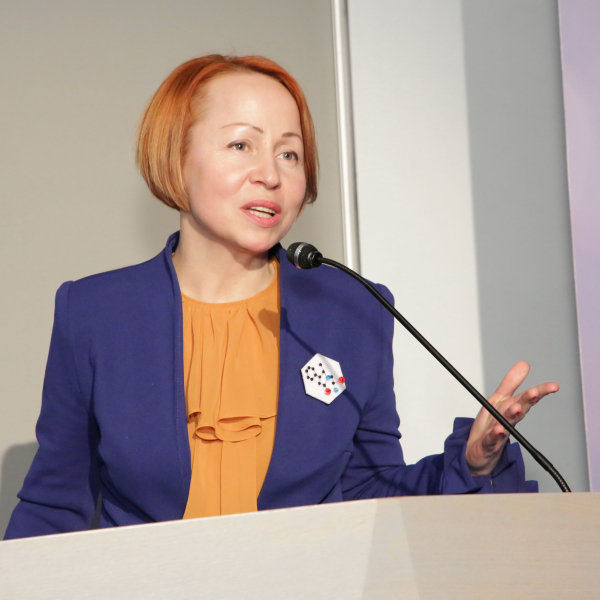
Dr. Liga Zvejniece, a Leading Researcher at the National Institute of Research and Innovation (NIRI) in Latvia, will explore experimental models of traumatic brain injury and their applications in biomedical research. Drawing on over two decades of experience in pharmacological research, she will discuss closed-skull models of brain trauma, their applications, and their relevance for understanding injury mechanisms and developing potential therapies.
This video is licensed under a Creative Commons Attribution-NonCommercial-NoDerivatives (CC BY-NC-ND) license. You are free to share it for non-commercial purposes with proper attribution, but no modifications or adaptations are allowed.
(Closed – skull injuries)
1. Why use this method?
Traumatic brain injury (TBI) is caused by external mechanical forces acting on the brain. Because human TBI is highly complex and heterogeneous, experimental animal models are necessary to better understand disease mechanisms and evaluate potential therapies.
The manual Shohami and Marmarou Weight Drop variants remain the most widely used, accessible, and foundational models for reproducing closed-skull TBI, which is why this protocol focuses specifically on their practical execution. These closed-skull injury models preserve the integrity of the skull to better mimic human concussion and diffuse traumatic brain injury. Within this framework, the Shohami model is utilized to mainly produce focal cortical injury, whereas the Marmarou model is applied to reproduce diffuse brain injury and diffuse axonal injury. Models are simple, inexpensive, relatively reproducible, and widely used to investigate traumatic brain injury.
While alternative closed-head methodologies exist, they carry distinct limitations; for instance, the automated CHIMERA system is often associated with high data variability, whereas the awake ACHI model presents significant regulatory challenges regarding ethical approvals. Other techniques, such as open-skull models requiring a craniectomy, fall entirely outside the scope of this procedure.
2. What you’ll need
What you will need to perform closed-skull injury
Category
Equipment / Material
Shohami Model
Marmarou Model
Animals
Mice or rats
✔
✔
Main Apparatus
Weight Drop apparatus
✔
✔
Model-Specific
Impact cone (tip diameter >5 mm)
✔
Model-Specific
Impact disk
✔
Model-Specific
Helmet disk (over the head)
✔
Surgical Tools
Surgical instruments
✔
✔
Preparation
Permanent marker
✔
✔
Preparation
Anaesthesia and Oxygen supply
✔
✔
Post-op Care
Polypropylene sutures (6-0 for mice or 4-0 for rats) or Hair clipper
✔
✔
Anaesthesia:
isoflurane (inhalation) or ketamine/xylazine (injectable)
Analgesia:
tramadol (10–25 mg/kg, s.c.) or buprenorphine (0.1 mg/kg, s.c.) or Meloxicam (1 mg/kg, s.c.)
Alcohol for surgical tool disinfection
Ophthalmic gel
3. Step-by-step instructions
Step 1. Ethical approval
Ensure that all experiments comply with national and institutional regulations regarding the use of animals for research purposes.
Step 2. Animal preparation
Handle the animals;
Weigh each animal;
Administer analgesia (tramadol, buprenorphine or meloxicam);
Induce and maintain anaesthesia using volatile isoflurane, or administer injectable anaesthetics (ketamine/xylazine or sodium pentobarbital);Protect the eyes using ophthalmic gel:
Trim the hair:
Disinfect the scalp with 70% alcohol.
Step 3. Surgical preparation
Make a midline longitudinal incision to expose the skull;
Visually identify the sagittal and coronal sutures;
Locate the impact area using a permanent marker;
Step 4. Weight Drop injury
Place the animal in the Weight Drop apparatus;
Place the helmet disk (Marmarou model) or position the impact cone (>5 mm) above the marked impact site (Shohami model);
Allow the calibrated weight to fall onto the impact site to induce traumatic brain injury.
Step 5. Post-operative care
In case of respiratory arrest more then 15 s, administer oxygen (3–5 L/min);
Close the scalp wound using polypropylene sutures: 6-0 for mice, 4-0 for rats;
Allow the animal to recover in a 37 °C heated recovery cage.
Step 6. Immediate behavioural characterization
Immediately after injury assess:
Duration of apnea (seconds);
Duration of loss of the righting reflex (seconds);
Presence of seizures.
Step 7. Neurological Severity Score (NSS)
Evaluate neurological deficits using the Neurological Severity Score.
The NSS consists of 10 behavioural tasks:
Exit circle – ability and initiative to exit a 30 cm diameter circle within 3 minutes;
Monoparesis/Hemiparesis – paresis of the contralateral upper and/or lower limb;
Straight walk – assessment of alertness, initiative and ability to walk in a straight line;
Startle reflex – response to a loud hand clap;
Seeking behaviour – physiological exploratory behaviour indicating interest in the environment;
Beam walk (3 cm) – ability to cross a 30 cm beam with a width of 3 cm;
Beam walk (2 cm) – same task performed on a 2 cm beam;
Beam walk (1 cm) – same task performed on a 1 cm beam;
Vertical beam balancing – ability to balance on a vertical beam (7 mm width) for at least 10 seconds;
Round stick balancing – ability to balance on a horizontal round stick (5 mm diameter) for at least 10 seconds;
Select the experimental model according to the scientific question being investigated;
Closed-skull models better mimic concussion and diffuse traumatic brain injury because the skull remains intact;
The Weight Drop model is technically simple and relatively inexpensive;
Injury severity may vary depending on impact positioning and impact conditions;
The operator must manually intercept the weight on its first rebound to avoid a second impact;
Double-check the exact calibrated height before every drop to maintain consistent energy transfer;
Exclusion criteria: Exclude any animal that sustains a skull fracture during the weight-drop procedure.
5. Critical appraisal & implications for future research
The Weight Drop model is one of the most commonly used experimental models of traumatic brain injury because it is simple, inexpensive, and relatively reproducible. The Shohami model mainly produces focal injury, whereas the Marmarou model better reproduces diffuse brain injury and diffuse axonal injury.
However, the model has several limitations. Injury severity may vary depending on impact position and impact conditions. Closed-skull models are generally associated with greater variability than open-skull models. Modified Weight Drop models improve clinical relevance but may differ between laboratories.
No single experimental model reproduces all aspects of human traumatic brain injury. Some models are better suited for focal cortical injury, whereas others more accurately reproduce diffuse axonal injury, concussion, repetitive mild TBI, or blast injury. Therefore, the choice of model should always depend on the scientific question and the pathological mechanisms under investigation.
References
Zvejniece L, Stelfa G, Vavers E, Kupats E, Kuka J, Svalbe B, Zvejniece B, Albert-Weissenberger C, Sirén AL, Plesnila N, Dambrova M. Skull Fractures Induce Neuroinflammation and Worsen Outcomes after Closed Head Injury in Mice. J Neurotrauma. 2020 Jan 15;37(2):295-304. doi: 10.1089/neu.2019.6524.
Albert-Weißenberger, C., Várrallyay, C., Raslan, F. et al. An experimental protocol for mimicking pathomechanisms of traumatic brain injury in mice. Exp & Trans Stroke Med 4, 1 (2012). https://doi.org/10.1186/2040-7378-4-1.
Flierl MA, Stahel PF, Beauchamp KM, Morgan SJ, Smith WR, Shohami E. Mouse closed head injury model induced by a weight-drop device. Nat Protoc. 2009;4(9):1328-37. doi: 10.1038/nprot.2009.148. Morales DM, Marklund N, Lebold D, Thompson HJ, Pitkanen A, Maxwell WL, Longhi L, Laurer H, Maegele M, Neugebauer E, Graham DI, Stocchetti N, McIntosh TK. Experimental models of traumatic brain injury: do we really need to build a better mousetrap? Neuroscience. 2005;136(4):971-89. doi: 10.1016/j.neuroscience.2005.08.030.
Marmarou A, Foda MA, van den Brink W, Campbell J, Kita H, Demetriadou K. A new model of diffuse brain injury in rats. Part I: Pathophysiology and biomechanics. J Neurosurg. 1994 Feb;80(2):291-300. doi: 10.3171/jns.1994.80.2.0291.
Shapira Y, Shohami E, Sidi A, Soffer D, Freeman S, Cotev S. Experimental closed head injury in rats: mechanical, pathophysiologic, and neurologic properties. Crit Care Med. 1988 Mar;16(3):258-65. doi: 10.1097/00003246-19880
This protocol is licensed under a Creative Commons Attribution-NonCommercial (CC BY-NC) license, allowing sharing and adaptation for non-commercial purposes with proper attribution.
Adding comments is only possible for registered users.
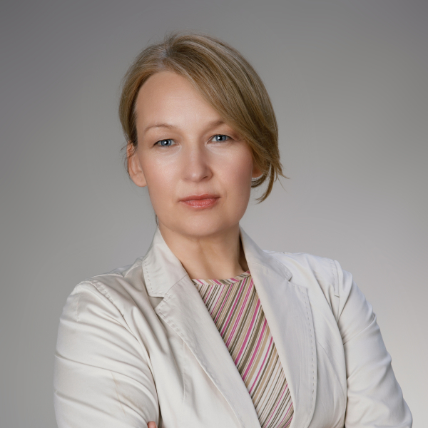
Dr. Liga Zvejniece is a Leading Researcher at the National Institute of Research and Innovation (NIRI) in Latvia. She has more than 20 years of experience in interdisciplinary pharmacological research, with a focus on neurodegenerative diseases, stroke, and traumatic brain injury. By utilizing behavioral tests, phenotyping of genetically modified animals, and biomedical/molecular biology approaches, her research centers on investigating the mechanisms of action of novel compounds. In addition to managing numerous academic research grants and European Regional Development Fund (ERDF) projects, Dr. Zvejniece has been responsible for animal protection and welfare at the institute since 2011. She also serves as a member of the Latvian Animal Protection Ethics Board and the National Committee for the protection of animals used for scientific purposes.






Adding comments is only possible for registered users.
Sign in