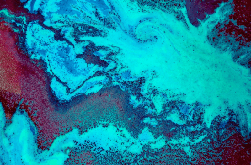
Rhinal cortex-hippocampus organotypic slices: A versatile model for epilepsy research
The rhinal cortex-hippocampus organotypic slice model provides a powerful platform for studying epilepsy by preserving the complex connectivity and architecture of critical brain regions. Dr. Cláudia Valente utilizes this model in conjunction with advanced imaging techniques, such as confocal microscopy and immunohistochemistry, to analyze neural circuits and molecular markers. This approach facilitates a deeper understanding of epileptogenesis and offers new insights into therapeutic strategies for epilepsy.
This video is licensed under a Creative Commons Attribution-NonCommercial-NoDerivatives (CC BY-NC-ND) license. You are free to share it for non-commercial purposes with proper attribution, but no modifications or adaptations are allowed.
1. Why use this method?
Organotypic slices from the rhinal cortex and hippocampus provide an ex vivo system to study epilepsy mechanisms. These slices preserve the complex connectivity and functional properties of the brain, allowing for detailed analysis of seizure-like activity, synaptic interactions, and the effects of pharmacological interventions. The model is especially advantageous for long-term experiments, enabling researchers to investigate the mechanisms of epileptogenesis over time.
2. What you’ll need
Materials and Equipment
Slice Preparation Tools:
- Dissecting microscope with bright illumination.
- Vibratome or Tissue chopper (optional, based on preference) for cutting precise slices (350 μm thickness).
- Sterile forceps and scalpels.
- Filter paper
Culture Medium Components:
- Dissection medium: 25 mM glucose in Gey’s Balanced Salt Solution (GBSS).
- Culture medium: 50% Opti-MEM, 25% HBSS, 25% Horse Serum (HS), 25 mM glucose, 30 μg/mL Gentamycin.
- Maintenance medium: Neurobasal-A (NBA), 2% B27, 1 mM L-glutamine, 30 μg/mL Gentamycin, HS (15%, 10%, 5% and 0%).
Incubation Setup:
- Humidified incubator set to 36-37°C with 5% CO₂.
Imaging Tools:
- Electrophysiology setup (if measuring neuronal activity).
- Epifluorescence or confocal microscope (for visualization of immunohistochemical images).
3. Step-by-step instructions
Step 1: Tissue Preparation
Medium preparation: Dissection medium: 25 mM glucose in Gey’s Balanced Salt Solution (GBSS). Culture medium: 50% Opti-MEM, 25% HBSS, 25% Horse Serum (HS), 25 mM glucose, 30 μg/mL Gentamycin. Maintenance medium: Neurobasal-A (NBA), 2% B27, 1 mM L-glutamine, 30 μg/mL Gentamycin, HS (15%, 10%, 5% and 0%).
- Dissect the brain from neonatal Sprague-Dawley rats postnatal 3-9 days (typically P6-P7) to maximize viability. Wash the brain 3 times in ice-cold GBSS.
- Under a dissecting microscope, keeping the brain in ice-cold GBSS, isolate each hemisphere and carefully remove the excess tissue that covers the hippocampus, without touching the hippocampal structure.
- Section the isolated tissue into slices (350 μm thick). Carefully separate the slices, keeping only the ones with a structurally intact rhinal cortex and hippocampus. DG and CA areas should be perfectly defined, as well as the entorhinal (EC) and perirhinal (PC) cortex.
- Place slices onto sterile culture inserts in a 6-well plate filled with culture medium.
Step 2: Culturing the Slices
- Incubate slices at 36-37°C in a humidified 5% CO₂ incubator.
- Change the medium every 2–3 days to maintain viability.
- Allow slices to stabilize for 7-14 days in vitro before experiments, enabling recovery and network development.
Step 3: Experimental Setup
- Cut the slice from the culture insert and transfer it to an interface chamber perfused with oxygenated culture medium for electrophysiological recordings.
- Use a syringe to fill the electrode with aCSF solution (NaCl, KCl, MgSO₄, NaHCO₃, glucose, and CaCl₂, bubbled with 95% O₂/5% CO₂) and insert glass electrode into receiving electrode.
- Wait until the temperature stabilizes at 37 °C.
- Record for 30 min with receiving electrode in CA3 area.
- Monitor spontaneous activity using field recordings (burst>10s, interburst interval>2s) and analyze using Clampfit software.
- For epilepsy research, MCC950, a small-molecule known for its specificity in targeting the NLRP3 inflammasome ameliorates the epileptic-like activity, reduced the protein levels of Iba-1 and GFAP, suggesting a decrease in microglia and astrocytes’ activation, affected microglia morphology promoting a shift in microglia phenotype and prevented Gasdermin D cleavage, pointing to a decrease in cell death by pyroptosis.
Step 4: Post-Experiment Analysis
- Fix slices in 4% paraformaldehyde for immunohistochemical analysis (primary and secondary antibodies application together with nuclear staining with Hoechst) or collect the tissue for Western Blot, ELISA or RT-PCR.
- Data can be analyzed using software tools, such as Clampfit for electrophysiological data and ImageJ or ZEN for immunohistochemical images.
4. Practical tips
Tissue Health: Use neonatal animals for better slice viability and ensure oxygenated solutions during preparation.
Connectivity: Verify under a microscope that slices maintain rhinal cortex-hippocampus connections.
Stability: Allow sufficient incubation time for recovery and network maturation before experiments.
Two methods were developed to maintain brain slices in a long-term culture:
– the roller tube technique, which has been shown to be difficult to prepare and known to bear a large experimental variability due to thinning of tissues;
– the membrane interface culture method, which maintains slices on a porous membrane at the interface between medium and a humidified atmosphere. The medium provides adequate nutrition to tissues through the membrane via capillary action.
In organotypic slices cell death can be easily assessed by monitoring the cellular uptake of the fluorescent dye propidium iodide (PI, 3,8-diamino-5-(3-(diethylmethylamino)propyl)-6-phenyl phenanthridinium diiodide).
PI is a polar compound which is taken up by cells with compromised membrane integrity, intercalates into DNA and emits red fluorescence (630 nm; absorbance 493 nm), which can be easily quantified using imaging software.
5. Critical appraisal & implications for future research
This model offers unparalleled access to the neural circuitry of the rhinal cortex and hippocampus, making it invaluable for epilepsy research. It enables precise control over experimental conditions while preserving connectivity and functionality. Future advancements in imaging techniques and molecular tools could further enhance the utility of this model, providing deeper insights into the mechanisms underlying epileptogenesis and potential therapeutic targets.
This protocol is licensed under a Creative Commons Attribution-NonCommercial (CC BY-NC) license, allowing sharing and adaptation for non-commercial purposes with proper attribution.


Adding comments is only possible for registered users.
Sign in